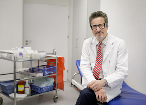
About „gold standards”, modern procedures and the future in neurosurgery

Patients with what diseases most often come to the Department of Neurosurgery of the Medical University of Warsaw?
In elective neurosurgery we have four main groups of patients: those with brain tumors, with brain aneurysms and hemangiomas, with spine pain and radiculalgia in the limbs, and people with functional disorders such as epilepsy. We also perform emergency procedures – this group are mostly patients after craniocerebral injuries.
What is your area of interest and what treatments do you specialize in?
I have developed and continue to develop in vascular neurosurgery, skull base surgery and minimally invasive spine surgery.
So let’s start with spine surgery. What did such procedures look like in the past and how are they done now?
When I became interested in neurosurgery over 30 years ago, all spine surgeries were “open”. No magnifying glasses or microscopes were used. At that time, in some hospitals, people after spine surgery were even recommended to stay in bed for two months. There is a myth that such a patient must avoid movement, or even vertical positioning, because it may cause a spine fracture. A lot has changed since then. It is easy to demonstrate on the example of our clinic. It was taken over by Prof. Andrzej Marchel in 1999 and he immediately introduced the use of a microscope in spine surgeries, which significantly reduced the invasiveness of the procedure. The incisions, which were previously about 10 centimeters long, became three or even four times shorter. A patient could go home after two or three days with just a small scar on their back. This minimally invasive procedure using a microscope, called “microdiscectomy”, is considered the “gold standard” to this day.
What about endoscopic procedures? Are they used in spine surgery?
Endoscopic spine procedures have been developing for several decades. However, the real boom occurred in the last decade. This is another revolution, but with a few caveats. It is worth remembering that not every patient is a good candidate for endoscopic surgery. Moreover, endoscopic techniques take quite a long time to learn. The neurosurgeon must “switch” from operating using a microscope (when he or she works with both hands) to a completely different situation in which the surgeon looks not towards the operating field on the patient, but at a computer display placed in the room. Moreover, he or she holds the endoscope in one hand, so there is only one hand left to perform all maneuvers. It takes practice. The acquisition of these skills is facilitated by the fact that endoscopic neurosurgery is developing not only in the field of spine surgery but also intracranial one. Therefore, this tool is increasingly used for various surgical purposes, and in the future most procedures will probably be performed in a minimally invasive manner, using endoscopes.
What spine problems can be solved endoscopically?
We use devices that allow us to remove pressure on the nerve roots and the spinal cord, as well as equipment that allows us to introduce various implants. A large part of spine procedures are so-called “stabilizations”. They are necessary when the patient has an unstable spine, for example because it was weakened during neurosurgery. In such cases, strengthening and stiffening the spine with implants or screws are needed to support the vertebrae in the gap after the removed disc. Most vendors of endoscopic sets for spine surgery also offer packages for inserting implants using this system. This means that we may perform almost all spine procedures endoscopically, at least in theory. However, as is the case with new products, there is still a critical evaluation of the results of endoscopic treatment ahead of us. It is worth remembering that “new” does not necessarily mean “better”. Spinal stabilization surgeries are very health-burdening procedures. For instance, about 60% of patients in Belgium cannot return to work after up to one year from spinal stabilization surgery, i.e. introducing screws into the spine.
Is this burden the same for both endoscopic and “open” surgeries?
The burden does not entirely depend on the technique. The treatment serves to achieve a specific goal. And the more difficult the goal and the more extensive the procedure, the greater the burden. Endoscopic surgeries have the main advantage that patients feel better and report less postoperative pain in the early period after surgery. In addition, a lower risk of infections has been demonstrated – this is due to the fact that there is less tissue damage, less blood loss, so the body is not so much weakened and the tissue exposure to infectious agents is lower.
What does it look like in the clinic you run – are spine surgeries more often performed “openly” or endoscopically?
When it comes to surgeries without the use of implants, i.e. simple strains or removal of a slipped disc, we performed the first endoscopic surgeries two years ago. However, I would like to emphasize that in our clinic, among various options, we choose the treatment that will achieve the best effect with the least invasive intervention possible. This means that we operate on many patients using a microscope. However, in the case of instrumental spine stabilization surgery, about 10 years ago I introduced a new technique into clinical practice in Poland – it is called “cortical screw trajectory”. This is a technique that allows much less interference in terms of access to the spine. Traditional stabilization involves spreading the spine muscles sideways – a bit like at a butcher’s. And this causes great trauma to the muscles, their denervation, then atrophy, and much more blood loss. The “cortical screw trajectory” procedure means that the same effect and scope of surgery can be achieved through a small, four-centimeter, incision. Starting from such a single incision, we can widen the spinal canal, remove the entire intervertebral disc, insert implants into the interbody space and, finally, drive screws into both adjacent vertebral bodies. In this way we achieve a complete decompression and stabilization effect without the need to injure the muscles. Two years ago we published a paper on the longest follow-up of patients after surgery performed using this technique. The conclusions are that this technique is as effective as traditional stabilization, and more beneficial for patients who recover faster. It is worth emphasizing that in the last two years there has been rapid progress in spine surgery. We have implemented a number of new techniques into our practice. We also introduce lateral and anterolateral approaches for stabilization of the lumbar spine, as well as percutaneous stabilization, which involves inserting screws in through incisions only to such a length that they can be screwed into the spine.
The clinic does brain tumor surgeries. Can such procedures be performed using endoscopic methods?
Neuro-oncology is our core scope of activity. We are known for this on the health services market. We have tradition, experience and continue to develop in the field of treatment of brain and spinal tumors. As for the surgical technique, the main trend is microsurgery, i.e. the use of a microscope and microtools. We use the endoscopic technique increasingly often, but only for surgery of intracranial tumors. This applies primarily to skull base surgery, i.e., for example, pituitary tumors, skull base bone tumors and skull base meningiomas. These procedures require cooperation not only of the neurosurgeon and anesthesiologist, but of the entire interdisciplinary team, so we cooperate with the Department of Otorhinolaryngology, Head and Neck Surgery, as well as with the Department of Internal Diseases and Endocrinology. These surgeries are most often performed by a team led by Dr. Tomasz Dziedzic and Dr. Tomasz Gotlib. These are so-called “four-handed procedures”: two surgeons operate and help each other. These surgeries are not only performed endoscopically – they are also supported by neuronavigation. Neuronavigation is a complicated process that allows the neurosurgeon see and control what he or she is doing, which is based on preoperative tests (MRI and CT). Thanks to neuronavigation, the safety of procedures and their precision have improved significantly – the operator knows and sees better where he or she is operating. When operating using old techniques, it often turned out that a piece of the tumor remained in the skull because it was simply not visible under microscopic visualization. Endoscopy combined with neuronavigation is much more accurate.
How are aneurysms treated today?
It is worth realizing that aneurysm is not a rare disease. It affects 3% to 7% of the population (it depends on the criteria). Twenty years ago we mainly treated ruptured aneurysms, i.e. patients in serious, life-threatening, conditions after hemorrhage. Many of them died without receiving treatment or even after successful clipping of the aneurysm due to complications following severe hemorrhage. Currently, 90% of aneurysm surgeries are performed using transvascular methods, i.e. embolization. We gain access either through the groin or through the radial artery of the forearm, then we pass a catheter through the vessels to the area around the aneurysm and we introduce various devices, such as implants, to close it. Transvascular treatment methods are developing rapidly. Medical companies producing coils, stents or various other devices for closing aneurysms introduce additional improvements every year to improve the safety and effectiveness of such procedures. In our clinic we have been using intra-aneurismal for years. This is conventional embolization. We also use stents, such as modern flow-diverting stents. They are designed in such a way that they allow blood to flow into small arteries – which is desirable because closing any of them could cause paralysis – and, at the same time, cause blood to clot in the aneurysm sac. It is worth mentioning one more novelty that we have been using since last year. These are devices that stop the flow of blood into the aneurysm. A very clever patent thanks to which we do not leave anything in the blood vessel itself, but only in the aneurysm. We introduce an implant into the neck part, and it opens up like an umbrella. It attaches to the basal part of the aneurysm and prevents blood from inflowing inside. As a result, the aneurysm clots and is treated with a single device. This is the simplest and shortest procedure (of course, for the patient who qualifies for it). Moreover, after this procedure there is no need to use many months of antiplatelet treatment that is necessary in the case of implanting stents. Of course, not all aneurysm surgeries are performed endovascularly. For example, we are developing the intracranial bypass method, which is unique in Poland. These are procedures that are difficult to learn. In our clinic we can perform extra- and intra-cranial bypasses. Thanks to this, we can bring blood to the brain from the extracranial artery to the artery behind the aneurysm’s outlet and close the aneurysm, which cannot be treated with any endovascular method.
Are hemangiomas also treated with endovascular methods?
I would like to emphasize that endovascular methods are not the only ones we use in the clinic. Progress in the treatment of cerebrovascular diseases also applies to microsurgical, i.e. open, surgery. Still, the best way to treat cerebral hemangiomas is to remove them using the microneurosurgical method. These treatments are becoming safer and are being improved. We have more and more options to protect the patient during such surgery. For example, we perform intraoperative angiography. During the procedure we can also check the patient’s nervous functions using neuromonitoring. There are situations when conventional surgery is simply the best choice and that is why we are also developing in this area.
What will be the direction of development in neurosurgery – reducing invasiveness, robotic surgeries, or maybe something else?
I am convinced that neurosurgery will wither in some areas and, at the same time, completely new paths will open up for it. It is known that neurosurgery will cease to be an important procedure in the treatment of gliomas in 10 or 20 years. These are infiltrating tumors that would be best treated without cutting them out because they cannot be safely and completely removed from the brain. Therefore, the treatment of gliomas is not about developing neurosurgery, but rather about developing basic science and understanding the molecular structure and nature of these tumors. Thanks to this, it will be possible to work on effective chemotherapy. The future of neurosurgery will be neurorestoration. We learned about it a few months ago thanks to Elon Musk. The idea is that by implanting a device into the brain, we can improve or restore the patient’s lost neurological functions. However, it is not about experiments that would increase the brain capacity of a healthy person. Neurorestoration will allow us to restore the functions that have been lost. Therefore, it is hope for patients paralyzed after a stroke or after a spine fracture with damage to the cervical cord.
How would such a neurorestoration work?
We are talking about the Brain Computer Interface (BCI). Let’s imagine a situation where the cerebral cortex looks and functions normally, but the neural pathway between it and one of the limbs is irreversibly damaged. Thanks to BCI, we place electrodes in the motor cortex and through them the computer learns the language that nerve cells communicate with – this is called “hacking” or “decoding” the brain. If the computer learns what the brain “thinks”, it is a very short way to restoring movement in the limb. There are various ideas on how to do this in practice. For example, a robotic glove can be used. Then the patient moves their hand, but it is actually the glove that moves. A person thinks about movement and the chip converts this thought into a robotic movement of the hand.
And what will be the role of the neurosurgeon here?
It will be important to properly and safely implant electrodes or other devices into the brain. However, engineers, mathematicians, IT specialists, as well as rehabilitators and neuropsychologists will have a much more important role to play. It’s fascinating to imagine what neurosurgery will look like in another quarter of a century. There have been some amazing changes over the last 25 years that I have been working. And, as we know, progress is happening faster and faster. What will happen in 10 or 20 years? I think it may exceed our wildest imaginations.
Interviewed by Iwona Kołakowska
Fot. Michał Teperek
Communication and Promotion Office