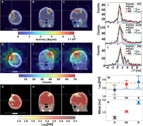
The study was designed to assess the feasibility of imaging cancerous lesions with a “positronium biomarker.” It was performed on a prototype J-PET apparatus - a novel tomograph developed and built by scientists from the Jagiellonian University. The prototype went to the Department of Nuclear Medicine UCC MUW, where specialists from this department performed the first imaging of a patient with a brain tumor.
An important part of the study was to compare the lifetime of positronium in healthy brain tissue and cancerous lesions, in this case glioma. The study showed that the average lifetime of positronium in glioma is significantly shorter than in healthy brain tissue. The findings suggest that positronium imaging could be used to increase the specificity of PET diagnostics in tissue pathology in vivo.
An article on the groundbreaking research has just been published in the prestigious journal Science Advances. It is available at the link
- Hopefully, this discovery will allow us to better diagnose patients with cancerous lesions in the future - says Prof. Jolanta Kunikowska.
As Prof. Kunikowska emphasized in an interview with MUW, currently specialists are already treating thyroid cancer, prostate cancer and neuroendocrine tumors with nuclear medicine methods. And ongoing research gives hope that many other cancers will soon be beaten in this way.
We encourage you to read the entire conversation available at the link
