The procedure was performed by the endoscopists - Jan Pertkiewicz, MD, PhD, and Slawomir Koziel, MD, in cooperation with the anesthesiology and nursing teams and representatives of the Boston Scientific company - the cholangioscope manufacturer.
In a patient, who underwent biliary reconstruction years ago after the damage during the cholecystectomy, the ductal-duodenal anastomosis narrowed late postoperatively and massive intrahepatic stones developed. The attempts to perform ERCP with a single-balloon enteroscope were unsuccessful for anatomical reasons. The patient underwent percutaneous bile duct drainage (PTCD) with balloon dilatation of the ductal-duodenal anastomosis and the placement of internal-external drainage. After several weeks, the drainage channel was widened and a SpyGlass Discover cholangioscope was inserted through this route. Bile ducts in both lobes of the liver were viewed confirming massive lithiasis. The stones were crushed into smaller fragments with the use of the electrohydraulic lithotripsy probe and generator. The basket and balloon were then used to evacuate the deposits and their fragments into the intestinal loop through the extanded anastomosis. The patient went home in very good general condition, free of aliments.
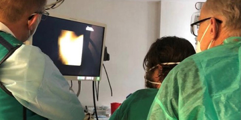
The SpyGlass Discover is a short, single-use endoscope used to explore the bile duct during the laparoscopic procedures or from the percutaneous access. Its unique design allows the tip to bend to 180º, which makes it possible to reach all branches of the biliary tree with a diameter of min 3mm. With high-resolution imaging, we can visualize even the most discrete changes in the bile-duct mucosa and the smallest deposits. A 1.2 mm working channel allows the insertion of biopsy forceps, electrohydraulic lithotripsy (EHL) or laser lithotripsy probes, tissue collection loops or cages for the evacuation of deposits. This allows the therapy to be performed under direct visual control, not just x-ray image, which greatly increases the effectiveness of the procedure and a patient safety. Currently, the Laboratory and Department’s team is in the final stage of purchasing the equipment used for this procedure, which will allow regular access to the described technique for other patients of our hospital.
Sławomir Kozieł, Krzysztof Zieniewicz


