Aortic valve stenosis is a narrowing of the lumen of the left arterial orifice resulting in obstructed outflow of blood from the left ventricle into the aorta. In the most severe forms, ventricular damage can result in the development of a single ventricle heart (HLHS) in the newborn and the need for multi-stage, extremely complicated surgery after birth. The diagnosis of fetal aortic valve stenosis is based on ultrasound examination of the fetal heart.
In the first patient, the surgery was performed at 23 weeks gestation. Doctors were able to safely dilate the aortic valve, and the baby has a chance to save the left ventricle. In case of the second patient, the situation was more complicated. The surgery was performed for vital indications because the child had circulatory failure and edema. He was threatened with death while still in the womb.
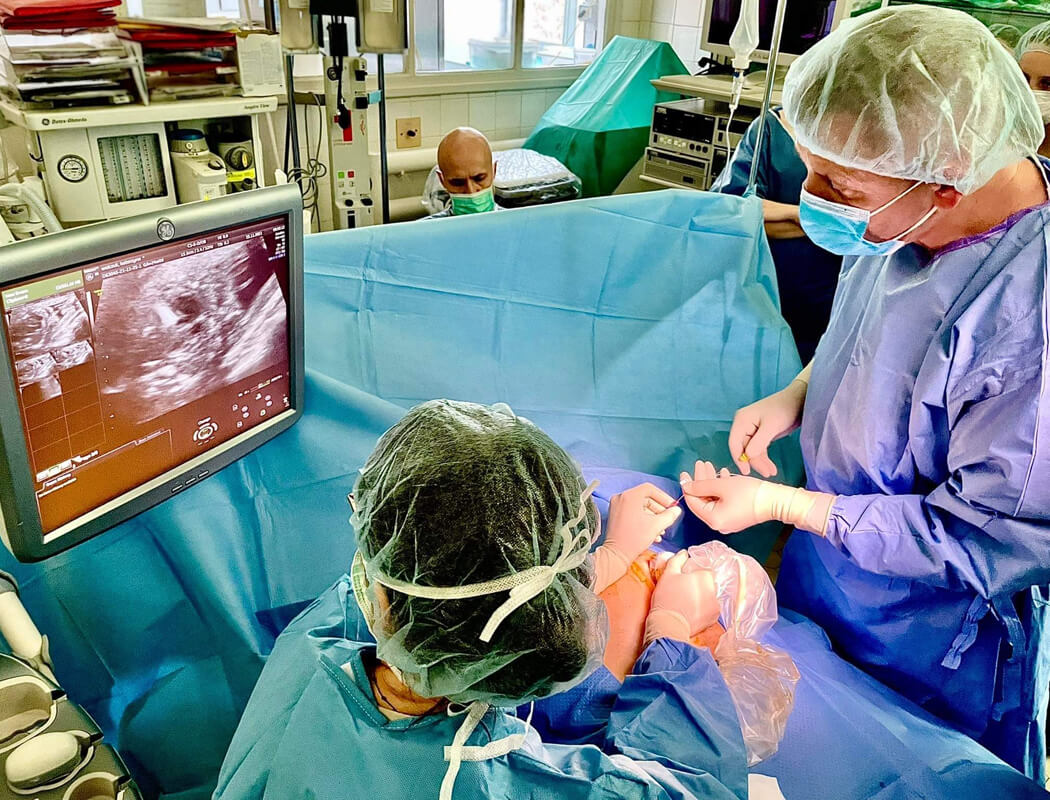
- We usually repair one little patient's heart in the womb in one day. Yesterday this occurred in two patients who were not yet born. There were difficult moments, but our Team showed that they can handle any situation. Now we keep our fingers crossed for good distant effects - says Professor Mirosław Wielgoś, head of the 1st Department of Obstetrics and Gynaecology MUW.
The surgeons who performed the operations: Prof. Marzena Dębska, Janusz Kochman, MD, PhD, Dr. Beata Rebizant and Dr. Katarzyna Zych-Krekora.