Microscopic structures of maximum importance

What exactly are extracellular vesicles?
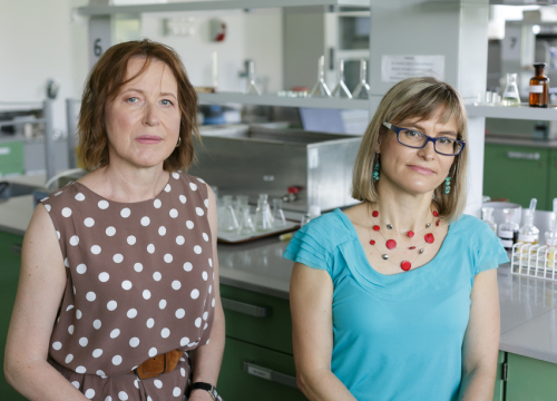
Dr. Małgorzata Czystowska-Kuźmicz: Extracellular vesicles (EVs) are microscopic structures with a double lipid membrane produced by all cells and, in particularly high concentrations, by cancer cells. It was once believed that they only play the role of “trash cans” for cells to get rid of harmful metabolic products or damaged organelles. However, numerous studies have shown in recent years that EVs play a key role in communication between cells, are necessary to maintain cellular homeostasis, and also significantly affect pathological processes, including the development of cancer. Because they are present in all biological fluids and reflect the molecular composition of the cells from which they originate, they are a potential source of biomarkers or therapeutic targets, also in the field of oncology. Currently, they are the subject of research around the world. They are also of interest to both start-ups in the field of biotechnology and large pharmaceutical companies. Thanks to intensive research, knowledge about them is growing very quickly, constantly creating new possibilities for their potential use in the clinic.
Why has such a “narrow” issue become the topic of the two-day conference?
Dr. Beata Pyrzyńska: The conference was designed to familiarize the scientific community at the Medical University of Warsaw and in Poland, especially students and young scientists, with the subject of extracellular vesicles and arouse their interest in this field of research. Foreign speakers, who are leading experts in the field of microvesicle research, as well as leading scientists from Poland, presented their latest studies in this field and summarized the latest knowledge on the subject. Their importance not only for oncology but also for regenerative medicine and a whole range of their potential clinical applications were discussed.
Which of the speeches and studies deserve a special mention?
Dr. Małgorzata Czystowska-Kuźmicz: The presence of Prof. Theresa L. Whiteside from the University of Pittsburgh, who is one of the pioneers of microvesicles research in immuno-oncology and has 35 years of experience in cancer immunology and monitoring the status of the immune system in cancer patients, was certainly of great importance. She was one of the first to demonstrate the effect of microvesicles of cancerous origin on the antitumor response of the immune system. She demonstrated at the conference that the molecular profile of exosomes (one of the vesicle subtypes) found in the plasma of cancer patients can be used to assess their immune status and allow to predict their response to immunotherapy. Interestingly, she showed that non-cancerous vesicles, e.g. originating from cells of the immune system, can be as much a source of diagnostic and prognostic biomarkers as cancerous vesicles. In addition, she presented various mechanisms used by exosomes to inhibit the antitumor response, emphasizing the need for a systemic approach when trying to block their immunosuppressive properties. Prof. Monika Pietrowska, a Polish expert in proteomic exosome research, cooperating with Prof. Whiteside, showed the options for using proteomics for molecular analysis of vesicles and proved that, with proper preparation of samples, it is an excellent method for identifying new diagnostic or prognostic markers associated with vesicles.
Dr. Beata Pyrzyńska: Another guest of the conference, Prof. Janusz Rak from McGill University in Montreal, the world-famous discoverer of oncogenic control of the processes of formation of vascularized tumor stroma, including the regulation of angiogenic factors, talked about his discovery of the spread of oncogenic proteins using microvesicles and their role in cancerous thrombosis. Prof. Rak emphasized that vesicles mediate not only the transfer of oncogenic proteins but also oncogenic mRNAs which initiate the protein gene expression in target cells, contributing to the pathological processes of angiogenesis and coagulation. Prof. Wei Guo from the University of Pennsylvania presented his latest research on the presence of various receptors, including the ICAM-1 protein, on the surface of extracellular vesicles of cancerous origin. He showed that, thanks to their presence, these vesicles can specifically “find” their targets, such as activated CD8+ T lymphocytes, and inhibit their functioning by interacting with the exosomal PD-L1 molecule with the PD-1 receptor on lymphocytes. Thus, the vesicles effectively eliminate effector lymphocytes crucial for the antitumor response, having a great impact on potential immunotherapies.
Prof. Tony Ng from King’s College of London argued that vesicles should be taken into account in the development of various platforms for stratification of patients in terms of risk, prognosis and therapies within the framework of personalized medicine. According to him, the molecular profile of vesicles can be as valuable a carrier of information as molecular imaging or analysis of the genetic or protein profile.
Dr. Małgorzata Czystowska-Kuźmicz: Prof. Bernd Giebel, President of the German Society for Extracellular Vesicles, an expert in the therapeutic use of microvesicles derived from mesenchymal stem cells, presented his latest research on standardizing the production of microvesicles for therapeutic purposes and their translation towards clinic applications, highlighting important problems that need to be addressed before vesicles are introduced into the clinic. Prof. Ewa Zuba-Surma, a Polish expert in the use of vesicles in regenerative medicine, explained the advantages of using vesicles for therapeutic purposes, compared to the use of stem cells alone. Prof. Joanna Rossowska presented her extremely ingenious ways of modifying vesicles of cancerous origin in order to increase their immunogenic abilities and, at the same time, reduce their suppressive potential. After such modification, these vesicles can be used as specific anti-cancer vaccines for dendritic cells.
Prof. Piotr Widłak drew attention to his discovery, extremely important for radiotherapy. The point is that vesicles mediate in inducing the so-called “bystander effect” in surrounding and distant tissues by ionizing radiation. They do this by inducing proteins associated with replication stress in target cells.
On the second day of the conference workshops on modern methods of isolation of extracellular vesicles and their characteristics were held. What is the problem with the isolation of EVs and why are the presented methods innovative?
Dr. Małgorzata Czystowska-Kuźmicz: The problem with the isolating and analyzing vesicles from biological fluids, i.e. the most clinically useful ones, is that the old, or “conventional”, methods do not provide sufficient separation of isolated vesicles from the accompanying protein impurities (e.g. albumin, plasma lipoprotein), which hinders and distorts subsequent analysis, especially proteomics. In addition, conventional insulation methods, such as ultracentrifugation, often damage the structure of vesicles and reduce their functionality.
Dr. Beata Pyrzyńska: The new methods of isolation and analysis presented at the workshop try to solve this problem. The method of isolation using the so-called size-exclusion chromatography allows us to get rid of most of the contaminating lipoproteins while isolating vesicles from plasma and while preserving their morphology and functionality. The participants first got acquainted with the theoretical basis of the method and then had the opportunity to carry out a practical exercise in isolating vesicles using this method from a real plasma sample taken from a patient. In terms of analysis, the conventional methods allowed only for aggregate analysis of the molecular composition of vesicles. Currently, under the influence of studies indicating the unusual diversity of microvesicles in biological samples, scientists suggest that a single-vesicle analysis is needed. Two of the latest analysis methods were presented during the workshop: nano-flow cytometry using Cytek’s Aurora cytometer and nanoparticle tracking analysis (NTA) with fluorescence using Particle Metrix’s Zetaview Quatt device. The participants analyzed their previously isolated microvesicles using the ZetaView instrument.
Conference agenda
Session 1: Extracellular vesicles in immuno-oncology and cancer research
- Prof. Theresa L. Whiteside, University of Pittsburgh, USA: “The potential role of exosomes from cancer plasma as biomarkers of cancer development”
- Prof. Monika Pietrowska, Maria Skłodowska-Curie National Institute of Oncology – National Research Institute, Gliwice Branch: “Small extracellular vesicles derived from melanoma cells – their role in cancer development”
- Prof. Janusz Rak, McGill University, Montreal, Canada: “The effect of oncogenes on regulatory signaling pathways associated with extracellular vesicles”
- Prof. Wei Guo, University of Pennsylvania, USA: “The importance of exosomes in immunosuppression and cancer development”
- Prof. Joanna Rossowska, Ludwik Hirszfeld Institute of Immunology and Experimental Therapy, Polish Academy of Sciences, Wrocław: “The role of modified exosomes of cancer origin in the modulation of the cancer microenvironment”
- Prof. Tony Ng, King’s College London, UK: “The use of exosomes as part of a comprehensive, multi-level strategy to stratify the risk of cancer patients in terms of disease progression in response to treatment”
Session 2: Extracellular vesicles in the clinic: therapeutic use, impact on cancer treatment
- Prof. Ewa Zuba-Surma, Jagiellonian University, Krakow: “Extracellular vesicles derived from stem cells as pro-regenerative regulators in tissue renewal”
- Prof. Bernd Giebel, University of Duisburg-Essen, Germany: "Clinical potential of extracellular vesicles derived from mesenchymal stem cells and challenges in their translational application”
- Prof. Piotr Widłak, Medical University of Gdańsk: “The bystander effect induced by ionizing radiation via extracellular vesicles is associated with replication stress in target cells.”
Session 3: Research related to extracellular vesicles as part of the EVIONA project at the Medical University of Warsaw
- Beata Pyrzyńska, Medical University of Warsaw: “Adverse effect of extracellular vesicles on the effectiveness of therapeutic antibodies”
- Karolina Soroczyńska, Medical University of Warsaw: “Detection of small extracellular vesicles containing arginase in biological fluids of endometriosis patients as potential immunosuppressive agents”
- Magdalena Długołęcka, Medical University of Warsaw: “Characterization of extracellular vesicles from bronchovesicular lavage fluid and from plasma of patients with lung lesions by nanoparticle tracking analysis”


