Evaluation of the effectiveness of methods for obtaining hypertrophy of liver parenchyma as a chance for patients with advanced liver cancers
Project titled "Comparison of the effectiveness of three different methods of obtaining liver parenchyma hypertrophy in patients qualified for partial resection of the organ (TANGO-LIVER Three Arm Nuclear Growth Observation in Liver Surgery)" received funding from the Medical Research Agency in the amount of PLN 4,556,510.76 as part of the competition for head-to-head research in the field of non-commercial clinical trials or research experiments (ABM/2022/3).
Why induce liver parenchymal hypertrophy?
Removal of a fragment of the liver remains the basic method of treatment for a large population of patients with primary and metastatic liver cancer. One of the most dangerous complications that can occur in the postoperative period is the failure of the remaining fragment. To put it simply - if we remove the "sick" part of the patient's liver, it may turn out that the "healthy" part left will be too small. In its most developed form, it leads to the death of the patient. Only in rare cases is it possible to perform a so-called emergency liver transplant. Due to the lack of effective causative treatment, one of the key elements of planning surgery is to determine the risk of postoperative liver failure.
Risk of postoperative liver failure
Among the numerous factors that influence this risk, the most important is the volume of the liver fragment planned for resection and coexisting damage to the parenchyma, for example during preoperative chemotherapy (which is very common in patients treated for cancer).
The recognized safety limit for partial hepatectomy is to leave a volume of parenchyma corresponding to 30% of the standard liver volume for a given patient. This value is adjusted to the quality of the parenchyma and should be higher in the case of significant fatty liver or fibrosis. If, at the stage of surgery planning, it turns out that the volume of the liver fragment to be left after resection is smaller than the established value, the tumors are considered initially unresectable. Most often, this leads to a situation where the only alternative is systemic palliative treatment, which has a huge adverse impact on long-term prognosis and survival chances.
Liver resection and the regenerative capacity of the liver
When planning liver resection, we can fortunately take advantage of the unique abilities of this unique organ, namely its ability to regenerate. This allows us to use one of several methods designed to induce hypertrophy of the part of the liver that will be left after surgery. However, this hypertrophy cannot affect the parts of the liver where the cancer foci are located, because it could result in uncontrolled spread of cancer cells and progression of the condition. Therefore, we must perform a procedure that will increase the volume of only the "healthy" part of the liver that we plan to leave in the patient.
Methods of inducing liver hypertrophy
World-recognized methods for inducing liver hypertrophy include: embolization of the portal vein branches (PVE) or embolization of the portal vein branches and hepatic veins (LVD) using interventional radiology techniques and surgical ligation of the portal branches with partial separation of the liver parenchyma (the so-called ALPPS operation – Associating Liver Partition and Portal vein Ligation for Staged hepatectomy).
In the first method (PVE), the portal blood flow to the "sick" part of the liver is blocked; in the second one (LVD), the venous outflow from the part of the liver is additionally closed. These procedures limit the blood flow to the fragment we want to remove, while compensatingly increasing blood flow to the fragment we want to leave. The third technique (ALPPS) involves surgical ligation of the portal vein branches and partial separation of the liver parenchyma between the fragment that is planned to be removed and the fragment that is planned to be left.
Previous experience shows that the effectiveness of achieving liver hypertrophy increases, but - unfortunately - with the risk of complications in all three methods: from radiological embolization of portal branches, through radiological embolization of portal branches and hepatic veins, to surgery using the ALPPS method. Unfortunately, there is currently no reliable scientific data to determine which method should be used in the first place. Existing research studies have been based on retrospective data, which is a significant limitation.
Evaluation of the effectiveness of hepatic hyperplasia
The effectiveness of achieving liver hypertrophy is currently assessed by calculating the volume of the organ fragment planned to be left on the basis of imaging tests performed, i.e. computed tomography or magnetic resonance imaging. Unfortunately, even with adequate liver volume, some patients develop liver failure in the postoperative period.
One of the important aspects that may be responsible for the development of this complication is the lack of functional assessment. It may provide information about functional reserve, which is crucial from the point of view of patient safety, in addition to the volume itself. In other words, we would like to predict not only the size of the fragment, but also its quality (function). Such an assessment, however, is not possible using only computed tomography. Therefore, we can use the scintigraphy test with 99mTc-mebrofenin, which allows us to estimate how well the fragment of the liver works by analyzing the clearance of mebrofenine. However, the test is currently not widely used due to much lower availability than computed tomography.
About the project
The planned study is intended to provide key information not only on the selection of the best method for obtaining liver parenchyma hypertrophy planned to be left, but will also compare methods allowing for the assessment of the effectiveness of inducing this hypertrophy. The study results will therefore translate not only into improved treatment outcomes, but also into greater patient safety.
The scientific project is to start in November 2023 and last until May 2029. The study will involve 154 patients with initially unresectable liver tumors. They will be randomly assigned to 3 different groups - in each group a different technique will be used to induce liver parenchyma hypertrophy (PVE, LVD or ALPPS). PAfter the procedure, patients will be monitored at weekly intervals using both CT scans and mebrofenine scintigraphy.
This will allow us to assess both the increase in the volume of a selected part of the liver and its function. All this to capture the moment when the remaining liver fragment reaches the functional capacity to take over the full functions of the liver. Thanks to this, it will be possible to carry out the next stage of treatment, i.e. liver resection, at the safest time for the patient.
For many patients, surgery after inducing liver hypertrophy is the only chance to cure cancer, which is why it is so important to conduct a study to show which method of inducing liver hypertrophy is the most effective and safe.
The results we obtain will allow us to plan the treatment of patients with liver cancer even better, and we hope that they will contribute to improving the results of treatment of patients in many centers around the world.
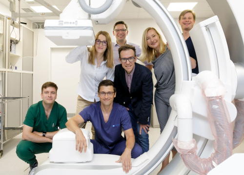
Clinical research team
The project team includes:
- Karolina Grąt, MD, PhD (project manager, specialist in radiology and imaging diagnostics),
- Krzysztof Korzeniowski, MD (interventional radiologist, specialist in radiology and imaging diagnostics),
- Krzysztof Lamparski, MD (interventional radiologist, specialist in radiology and imaging diagnostics),
- Professor Jolanta Kunikowska (specialist in nuclear medicine and internal medicine),
- Kacper Pełka, MD (nuclear medicine resident),
- Łukasz Masior, MD, PhD (specialist in general and oncological surgery),
- and Professor Michał Grąt (specialist in general surgery, oncology and clinical transplantology).
Editor: Communication and Promotion Office of the MUW
Fot. Michał Teperek; Communication and Promotion Office of the MUW