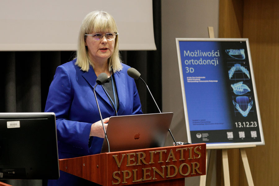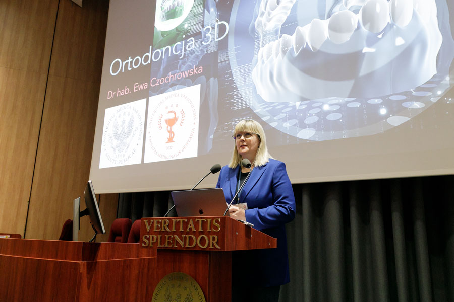
Beginning the lecture, the speaker emphasized the importance of the face, its aesthetics and smile in interpersonal communication and perception of the other person. She then presented the use of three-dimensional imaging in orthodontic and interdisciplinary planning and treatment in other areas of dentistry.
Orthodontics and aesthetics
- In orthodontics, we don't say whether the face is beautiful or ugly - said Ewa Czochrowska, MD, PhD. - When a patient comes to me, I first assess the symmetry and proportions of his face, because existing disorders most often reflect existing malocclusion.
And she presented selected clinical cases that were helped by orthodontic intervention. And not only in terms of improving oral health, but also in improving the wellbeing related to appearance or functional disorders. She spoke about a patient with significant protrusion and lateral displacement of the mandible, whose facial features improved very much after mandibular retraction surgery. She also presented the case of a patient who underwent orthognathic surgery for an aggravated open bite and lack of contact between teeth.
The vice dean also discussed cases of patients who undergo bite correction using tooth extractions (mainly first and second premolars). This is done when the patient's teeth are too large to fit within the jaws and are very crowded. As the speaker emphasized, jaw widening can be considered in such cases, but only to a certain extent, since the width of the face is not determined only by the jaw, but also, for example, by the zygomatic arch, the cheek muscles and the dimensions of the cerebral part of the skull.
- If teeth crowding is significant, teeth extractions are necessary to realign the front teeth, explained Ewa Czochrowska, MD, PhD.
Teeth transplants
The speaker presented the students with the case of a child who knocked out his upper central incisor and broke the root in the other. Implants were not an option for him, as this type of tooth replacement does not "follow" the growth of the jaw and face. Teeth autotransplantation proved to be a lifesaver. Two lower premolar teeth were transplanted in place of his lost incisors. The shape of the transplanted teeth was reshaped with composite material, the kind used for fillings in teeth. And, it is worth noting, the transplanted teeth behave just like the natural ones and can be moved with braces.
- The method of teeth autotransplantation is effective for teeth with undeveloped roots or teeth with few roots. It can be compared to planting trees: those with young roots are easier to transplant than those with well-developed, strong roots - explained Ewa Czochrowska, MD, PhD. And she emphasized: - The chances of transplanting such teeth are above 90%, i.e. it is successful in nine out of ten patients. Of course, the planning of the procedure is conducted on the basis of the analysis of the three-dimensional X-ray, so not every patient is qualified for this method. But those who are, have a very good chance of success of the procedure.
3D imaging in orthodontics
During the lecture, Ewa Czochrowska, MD, PhD, repeatedly spoke about improving the treatment process thanks to modern methods used in dentistry, primarily 3D imaging. Examples? Today, the place of the traditional plaster impression is being replaced by scanning the dental arches and creating digital models. This makes it possible to process a digital image of the patient's bite using various computer programs, and to prepare, for example, a dental crown and later print it. Or, for example, offer treatment with trays, changed every 7 to 14 days, which move the teeth.
The speaker also spoke about the use of extraoral scanners that memorize facial features objectively. Superimposing such scans on a 3D X-ray allows oral and maxillofacial surgeons and orthodontists to simulate bone displacements for orthognathic surgery and observe how the face might change after surgery.
Ewa Czochrowska, MD, PhD, finally stressed to remember that in treatment the most important thing is always the patient.
- We can have great technologies, but at the end of the treatment process we always have the patient and we need to understand what they want and what their expectations are - she stressed. And she added: - Undoubtedly, however, three-dimensional imaging technologies in orthodontics allow for better communication with the patient regarding treatment options and results, which helps in making an informed decision about the choice of dental therapy.
After the lecture, the students asked the speaker many questions about selected aspects of orthodontic treatment and the studies in the medical and dental field.
The meeting was attended by Klementyna Hoffmanowa High School No. IX, Władysław IV High School No. VIII, Stefan Batory High School No. II with Bilingual Branches, Tadeusz Czacki High School No. XXVII, Stefan Zeromski High School No. XL with Bilingual Branches.